Dental x-rays are photographs of the teeth and mouth. X-rays are a kind of electromagnetic radiation with high energy. The x-rays enter the body and create a picture on film or screen. Dental x-rays can be produced digitally or on film.
Dense structures (such as silver fillings or metal repairs) will block most of the x-ray’s light radiation. As a result, they seem white in the photograph. Structures containing air will be black, while teeth, tissue, and fluid will be grey.
Why are Dental X-Rays Performed?
Dental X-rays are usually taken once a year, and they may occur more frequently if your dentist monitors the development of a dental condition or therapy. Your age, your current oral health, any oral disease symptoms, a history of gum disease (gingivitis), or tooth decay factors may be among the factors that influence how frequently you have dental X-rays.
If you’re a new patient, you’ll almost certainly be exposed to dental X-rays for your new dentist to have a thorough picture of your oral health. Because their dentists may need to observe the growth of their adult teeth, children may require more regular dental X-rays than adults. This is crucial because it can help the dentist determine if baby teeth should be removed to avoid problems like adult teeth growing behind baby teeth.
People might ask, “Are dental x-rays safe?”. It is understandable if you have never had any experience with x-rays, whether dental or not, and the answer is it is entirely safe. If you are still concerned about it, you can check out the process of dental x-rays from this article.
Risks of Dental X-Rays
While dental X-rays do expose patients to radiation, the amounts are so minimal that they are deemed safe for both children and adults. Your radiation exposure risks are reduced even further if your dentist utilises digital X-rays instead of processing them on film.
Your dentist will also drape a lead “bib” over your chest, belly, and pelvic region to protect your essential organs from unwanted radiation exposure. A thyroid collar may be used to treat thyroid problems, and these may be worn in addition to the lead bib by children and women of reproductive age.
The exception to the rule is dental x-rays while pregnant. If you suspect you are pregnant, notify your dentist because radiation is unsafe for developing fetuses.
Preparing for Dental X-Rays
No specific preparation is required for dental X-rays. The only thing you’ll want to do before your visit is washing your teeth, creating a more hygienic atmosphere for those who work within your mouth. X-rays are typically taken before cleanings.
Types of X-Rays
There are wide varieties of dental X-rays, each capturing a somewhat different perspective of your mouth. Intraoral X-rays, for example, are the most prevalent. The list of dental x-rays can be found below.
- Bitewing
Your dentist can evaluate how well the crowns of your teeth line up by having you bite down on a certain piece of paper. This method is commonly used to discover cavities between teeth (interdental).
- Occlusal
When your jaw is closed, this X-ray examines how your upper and lower teeth line up. It can also identify anatomical anomalies in the mouth floor or palate.
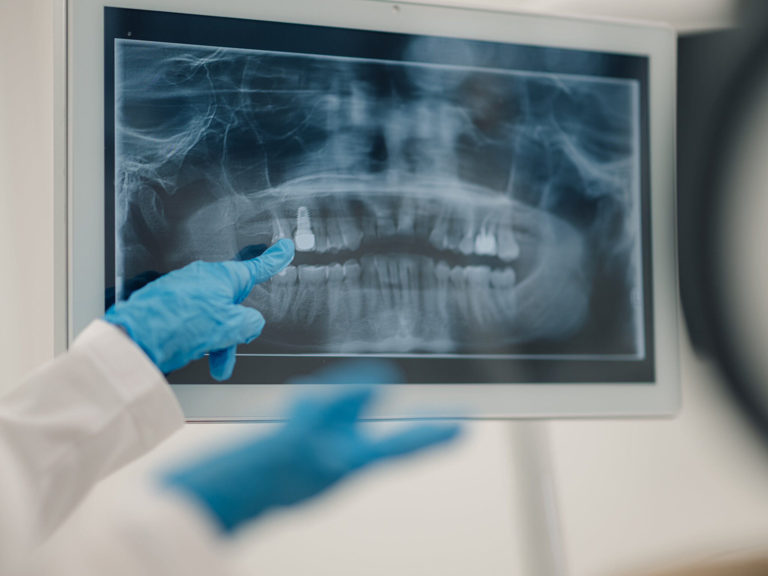
- Panoramic
For this type of X-ray, the machine revolves around the head. Your dentist might employ this technique to check your wisdom teeth, make arrangements for dental implants, or assess jaw issues.
- Periapical
This method concentrates on two whole teeth, from root to crown.
- Extraoral
These types of x-rays may be utilised when your dentist feels that there may be abnormalities in places other than the gums and teeth, such as the jaw.
A dental hygienist will guide each step of the X-ray procedure. They may leave the room temporarily while the photographs are being taken, and you will be told to remain still while the pictures are taken. If spacers (film holders) are utilised, they will be moved and adjusted in your mouth to acquire the correct photographs.
How is the Procedure Performed?
At the dentist, you’ll sit on a chair while wearing a lead vest across your chest and lap. The X-ray machine is placed near your head to photograph your mouth. While some dental offices maintain X-rays in a separate area from cleanings and other operations, others keep them in the same space.
After Dental X-Rays
When the photos are available — in the case of digital X-rays, quickly — your dentist will study them and look for any anomalies. If a dental hygienist cleans your teeth, the dentist may review the X-ray results with you after the cleaning. The hygienist may make an exception if major concerns are discovered during the X-rays. If your dentist detects any problems, such as cavities or tooth rot, they will discuss your treatment choices.
You can read our previous article from https://smileteamturkey.com/blog/diabetes-and-oral-health/.